Meiosis
| It has been suggested that Origin and function of meiosis be merged into this article or section. (Discuss) |

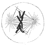
Before meiosis, the cell's chromosomes are duplicated by a round of DNA replication. This leaves the maternal and paternal versions of each chromosome, called homologs, composed of two exact copies called sister chromatids and attached at the centromere region. In the beginning of meiosis, the maternal and paternal homologs pair to each other. Then they typically exchange parts by homologous recombination, leading to crossovers of DNA from the maternal version of the chromosome to the paternal version and vice versa. Spindle fibers bind to the centromeres of each pair of homologs and arrange the pairs at the spindle equator. Then the fibers pull the recombined homologs to opposite poles of the cell. As the chromosomes move away from the center, the cell divides into two daughter cells, each containing a haploid number of chromosomes composed of two chromatids. After the recombined maternal and paternal homologs have separated into the two daughter cells, a second round of cell division occurs. There, meiosis ends as the two sister chromatids making up each homolog are separated and move into one of the four resulting gamete cells. Upon fertilization, for example when a sperm enters an egg cell, two gamete cells produced by meiosis fuse. The gamete from the mother and the gamete from the father each contribute half to the set of chromosomes that make up the new offsping's genome.
Meiosis uses many of the same mechanisms as mitosis, a type of cell division used by eukaryotes like plants and animals to split one cell into two identical daughter cells. In all plants, and in many protists, meiosis results in the formation of spores, haploid cells that can divide vegetatively without undergoing fertilization. Some eukaryotes, like Bdelloid rotifers, have lost the ability to carry out meiosis and have acquired the ability to reproduce by parthenogenesis. Meiosis does not occur in archaea or bacteria, which reproduce via asexual processes such as binary fission.
Contents[hide] |
[edit] History
Meiosis was discovered and described for the first time in sea urchin eggs in 1876 by the German biologist Oscar Hertwig. It was described again in 1883, at the level of chromosomes, by the Belgian zoologist Edouard Van Beneden, in Ascaris worms' eggs. The significance of meiosis for reproduction and inheritance, however, was described only in 1890 by German biologist August Weismann, who noted that two cell divisions were necessary to transform one diploid cell into four haploid cells if the number of chromosomes had to be maintained. In 1911, the American geneticist Thomas Hunt Morgan observed crossover in Drosophila melanogaster meiosis and provided the first genetic evidence that genes are transmitted on chromosomes. The term meiosis was coined by J.B Farmer and J.B Moore in 1905.[edit] Evolution
Meiosis is thought to have appeared 1.4 billion years ago. The only supergroup of eukaryotes which does not have meiosis in all organisms is excavata. The other five major supergroups, opisthokonts, amoebozoa, rhizaria, archaeplastida and chromalveolates all seem to have genes for meiosis universally present, even if not always functional. Some excavata species do have meiosis which is consistent with the hypothesis that this group is an ancient, paraphyletic grade. An example of a eukaryotic organism in which meiosis does not exist is euglenoid.[edit] Occurrence of meiosis in eukaryotic life cycles
Cycling meiosis and fertilization events produces a series of transitions back and forth between alternating haploid and diploid states. The organism phase of the life cycle can occur either during the diploid state (gametic or diploid life cycle), during the haploid state (zygotic or haploid life cycle), or both (sporic or haplodiploid life cycle, in which there two distinct organism phases, one during the haploid state and the other during the diploid state). In this sense, there are three types of life cycles that utilize sexual reproduction, differentiated by the location of the organisms phase(s).
In the gametic life cycle, of which humans are a part, the species is diploid, grown from a diploid cell called the zygote. The organism's diploid germ-line stem cells undergo meiosis to create haploid gametes (the spermatozoa for males and ova for females), which fertilize to form the zygote. The diploid zygote undergoes repeated cellular division by mitosis to grow into the organism. Mitosis is a related process to meiosis that creates two cells that are genetically identical to the parent cell. The general principle is that mitosis creates somatic cells and meiosis creates germ cells.
In the zygotic life cycle the species is haploid instead, spawned by the proliferation and differentiation of a single haploid cell called the gamete. Two organisms of opposing gender contribute their haploid germ cells to form a diploid zygote. The zygote undergoes meiosis immediately, creating four haploid cells. These cells undergo mitosis to create the organism. Many fungi and many protozoa are members of the zygotic life cycle.
Finally, in the sporic life cycle, the living organism alternates between haploid and diploid states. Consequently, this cycle is also known as the alternation of generations. The diploid organism's germ-line cells undergo meiosis to produce spores. The spores proliferate by mitosis, growing into a haploid organism. The haploid organism's germ cells then combine with another haploid organism's cells, creating the zygote. The zygote undergoes repeated mitosis and differentiation to become the diploid organism again. The sporic life cycle can be considered a fusion of the gametic and zygotic life cycles.
[edit] Process
Because meiosis is a "one-way" process, it cannot be said to engage in a cell cycle as mitosis does. However, the preparatory steps that lead up to meiosis are identical in pattern and name to the interphase of the mitotic cell cycle.Interphase is divided into three phases:
- Growth 1 (G1) phase: This is a very active period, where the cell synthesizes its vast array of proteins, including the enzymes and structural proteins it will need for growth. In G1 stage each of the chromosomes consists of a single (very long) molecule of DNA. In humans, at this point cells are 46 chromosomes, 2N, identical to somatic cells.
- Synthesis (S) phase: The genetic material is replicated: each of its chromosomes duplicates, so that each of the 46 chromosomes becomes a complex of two identical sister chromatids. The cell is still considered diploid because it still contains the same number of centromeres. The identical sister chromatids have not yet condensed into the densely packaged chromosomes visible with the light microscope. This will take place during prophase I in meiosis.
- Growth 2 (G2) phase: G2 phase is absent in Meiosis
Meiosis II consists of decoupling each chromosome's sister strands (chromatids), and segregating the individual chromatids into haploid daughter cells. The two cells resulting from meiosis I divide during meiosis II, creating 4 haploid daughter cells. Meiosis I and II are each divided into prophase, metaphase, anaphase, and telophase stages, similar in purpose to their analogous subphases in the mitotic cell cycle. Therefore, meiosis includes the stages of meiosis I (prophase I, metaphase I, anaphase I, telophase I), and meiosis II (prophase II, metaphase II, anaphase II, telophase II).
Meiosis generates genetic diversity in two ways: (1) independent alignment and subsequent separation of homologous chromosome pairs during the first meiotic division allows a random and independent selection of each chromosome segregates into each gamete; and (2) physical exchange of homologous chromosomal regions by homologous recombination during prophase I results in new combinations of DNA within chromosomes.
[edit] Phases
Meiosis takes place in several stages.[edit] Meiosis I
Meiosis I separates homologous chromosomes, producing two haploid cells (N chromosomes, 23 in humans), so meiosis I is referred to as a reductional division. A regular diploid human cell contains 46 chromosomes and is considered 2N because it contains 23 pairs of homologous chromosomes. However, after meiosis I, although the cell contains 46 chromatids, it is only considered as being N, with 23 chromosomes. This is because later, in Anaphase I, the sister chromatids will remain together as the spindle fibres pulls the pair toward the pole of the new cell. In meiosis II, an equational division similar to mitosis will occur whereby the sister chromatids are finally split, creating a total of 4 haploid cells (23 chromosomes, N) per daughter cell from the first division.[edit] Prophase I
During prophase I, DNA is exchanged between homologous chromosomes in a process called homologous recombination. This often results in chromosomal crossover. The new combinations of DNA created during crossover are a significant source of genetic variation, and may result in beneficial new combinations of alleles. The paired and replicated chromosomes are called bivalents or tetrads, which have two chromosomes and four chromatids, with one chromosome coming from each parent. At this stage, non-sister chromatids may cross-over at points called chiasmata (plural; singular chiasma).[edit] Leptotene
The first stage of prophase I is the leptotene stage, also known as leptonema, from Greek words meaning "thin threads".[1]:27In this stage of prophase I, individual chromosomes—each consisting of two sister chromatids—change from the diffuse state they exist in during the cell's period of growth and gene expression, and condense into visible strands within the nucleus.[1]:27[2]:353 However the two sister chromatids are still so tightly bound that they are indistinguishable from one another. During leptotene, lateral elements of the synaptonemal complex assemble.Leptotene is of very short duration and progressive condensation and coiling of chromosome fibers takes place. Chromosome assume a long thread like shape,they contract and become thick.At the beginning chromosomes are present in diploid number as in mitotic prophase.Each chromosome is made up of only one chromosome and half of the total chromosome are paternal and half maternal.For every paternal chromosome there is a corresponding maternal chromosome similar in size,shape and nature of inherited characters and are called homologous chromosome.[edit] Zygotene
The zygotene stage, also known as zygonema, from Greek words meaning "paired threads",[1]:27 occurs as the chromosomes approximately line up with each other into homologous chromosome pairs. This is called the bouquet stage because of the way the telomeres cluster at one end of the nucleus. At this stage, the synapsis (pairing/coming together) of homologous chromosomes takes place, facilitated by assembly of central element of the synaptonemal complex.Pairing is brought about by a zipper like fashion and may start at the centromere(procentric),at the chromosome ends(proterminal),or at any other portion(intermediate).Individuals of a pair are equal in length and in position of centromere. Thus pairing is highly specific and exact.The paired chromosomes are called Bivalent chromosome.[edit] Pachytene
The pachytene stage, also known as pachynema, from Greek words meaning "thick threads",[1]:27 is the stage when chromosomal crossover (crossing over) occurs. Nonsister chromatids of homologous chromosomes randomly exchange segments over regions of homology. Sex chromosomes, however, are not wholly identical, and only exchange information over a small region of homology. At the sites where exchange happens, chiasmata form. The exchange of information between the non-sister chromatids results in a recombination of information; each chromosome has the complete set of information it had before, and there are no gaps formed as a result of the process. Because the chromosomes cannot be distinguished in the synaptonemal complex, the actual act of crossing over is not perceivable through the microscope, and chiasmata are not visible until the next stage.[edit] Diplotene
During the diplotene stage, also known as diplonema, from Greek words meaning "two threads",[1]:30 the synaptonemal complex degrades and homologous chromosomes separate from one another a little. The chromosomes themselves uncoil a bit, allowing some transcription of DNA. However, the homologous chromosomes of each bivalent remain tightly bound at chiasmata, the regions where crossing-over occurred. The chiasmata remain on the chromosomes until they are severed in anaphase I.In human fetal oogenesis all developing oocytes develop to this stage and stop before birth. This suspended state is referred to as the dictyotene stage and remains so until puberty. In males, only spermatogonia (spermatogenesis) exist until meiosis begins at puberty.
[edit] Diakinesis
Chromosomes condense further during the diakinesis stage, from Greek words meaning "moving through".[1]:30 This is the first point in meiosis where the four parts of the tetrads are actually visible. Sites of crossing over entangle together, effectively overlapping, making chiasmata clearly visible. Other than this observation, the rest of the stage closely resembles prometaphase of mitosis; the nucleoli disappear, the nuclear membrane disintegrates into vesicles, and the meiotic spindle begins to form.[edit] Synchronous processes
During these stages, two centrosomes, containing a pair of centrioles in animal cells, migrate to the two poles of the cell. These centrosomes, which were duplicated during S-phase, function as microtubule organizing centers nucleating microtubules, which are essentially cellular ropes and poles. The microtubules invade the nuclear region after the nuclear envelope disintegrates, attaching to the chromosomes at the kinetochore. The kinetochore functions as a motor, pulling the chromosome along the attached microtubule toward the originating centriole, like a train on a track. There are four kinetochores on each tetrad, but the pair of kinetochores on each sister chromatid fuses and functions as a unit during meiosis I.[3][4]Microtubules that attach to the kinetochores are known as kinetochore microtubules. Other microtubules will interact with microtubules from the opposite centriole: these are called nonkinetochore microtubules or polar microtubules. A third type of microtubules, the aster microtubules, radiates from the centrosome into the cytoplasm or contacts components of the membrane skeleton.
[edit] Metaphase I
Homologous pairs move together along the metaphase plate: As kinetochore microtubules from both centrioles attach to their respective kinetochores, the homologous chromosomes align along an equatorial plane that bisects the spindle, due to continuous counterbalancing forces exerted on the bivalents by the microtubules emanating from the two kinetochores of homologous chromosomes. The physical basis of the independent assortment of chromosomes is the random orientation of each bivalent along the metaphase plate, with respect to the orientation of the other bivalents along the same equatorial line.[edit] Anaphase I
Kinetochore (bipolar spindles) microtubules shorten, severing the recombination nodules and pulling homologous chromosomes apart. Since each chromosome has only one functional unit of a pair of kinetochores,[4] whole chromosomes are pulled toward opposing poles, forming two haploid sets. Each chromosome still contains a pair of sister chromatids. Nonkinetochore microtubules lengthen, pushing the centrioles farther apart. The cell elongates in preparation for division down the center.[edit] Telophase I
The last meiotic division effectively ends when the chromosomes arrive at the poles. Each daughter cell now has half the number of chromosomes but each chromosome consists of a pair of chromatids. The microtubules that make up the spindle network disappear, and a new nuclear membrane surrounds each haploid set. The chromosomes uncoil back into chromatin. Cytokinesis, the pinching of the cell membrane in animal cells or the formation of the cell wall in plant cells, occurs, completing the creation of two daughter cells. Sister chromatids remain attached during telophase I.Cells may enter a period of rest known as interkinesis or interphase II. No DNA replication occurs during this stage.
[edit] Meiosis II
Meiosis II is the second part of the meiotic process. Much of the process is similar to mitosis. The end result is production of four haploid cells (23 chromosomes, 1N in humans) from the two haploid cells (23 chromosomes, 1N * each of the chromosomes consisting of two sister chromatids) produced in meiosis I. The four main steps of Meiosis II are: Prophase II, Metaphase II, Anaphase II, and Telophase II.In prophase II we see the disappearance of the nucleoli and the nuclear envelope again as well as the shortening and thickening of the chromatids. Centrioles move to the polar regions and arrange spindle fibers for the second meiotic division.
In metaphase II, the centromeres contain two kinetochores that attach to spindle fibers from the centrosomes (centrioles) at each pole. The new equatorial metaphase plate is rotated by 90 degrees when compared to meiosis I, perpendicular to the previous plate[citation needed].
This is followed by anaphase II, where the centromeres are cleaved, allowing microtubules attached to the kinetochores to pull the sister chromatids apart. The sister chromatids by convention are now called sister chromosomes as they move toward opposing poles.
The process ends with telophase II, which is similar to telophase I, and is marked by uncoiling and lengthening of the chromosomes and the disappearance of the spindle. Nuclear envelopes reform and cleavage or cell wall formation eventually produces a total of four daughter cells, each with a haploid set of chromosomes. Meiosis is now complete and ends up with four new daughter cells.
[edit] Significance
Meiosis facilitates stable sexual reproduction. Without the halving of ploidy, or chromosome count, fertilization would result in zygotes that have twice the number of chromosomes as the zygotes from the previous generation. Successive generations would have an exponential increase in chromosome count. In organisms that are normally diploid, polyploidy, the state of having three or more sets of chromosomes, results in extreme developmental abnormalities or lethality.[5] Polyploidy is poorly tolerated in most animal species. Plants, however, regularly produce fertile, viable polyploids. Polyploidy has been implicated as an important mechanism in plant speciation.Most importantly, recombination and independent assortment of homologous chromosomes allow for a greater diversity of genotypes in the offspring. This produces genetic variation in gametes that promote genetic and phenotypic variation in a population of offspring. Therefore a gene for meiosis will be favoured by natural selection over an allele for mitotic reproduction, because any selection pressure which acts against any clone will act against all clones, whilst inevitably favoring some offspring which are the result of sexual reproduction.
[edit] Nondisjunction
This is a cause of several medical conditions in humans (such as):
- Down Syndrome - trisomy of chromosome 21
- Patau Syndrome - trisomy of chromosome 13
- Edward Syndrome - trisomy of chromosome 18
- Klinefelter Syndrome - extra X chromosomes in males - i.e. XXY, XXXY, XXXXY
- Turner Syndrome - lacking of one X chromosome in females - i.e. XO
- Triple X syndrome - an extra X chromosome in females
- XYY Syndrome - an extra Y chromosome in males
[edit] Meiosis in mammals
In females, meiosis occurs in cells known as oogonia (singular: oogonium). Each oogonium that initiates meiosis will divide twice to form a single oocyte and two polar bodies.[6] However, before these divisions occur, these cells stop at the diplotene stage of meiosis I and lie dormant within a protective shell of somatic cells called the follicle. Follicles begin growth at a steady pace in a process known as folliculogenesis, and a small number enter the menstrual cycle. Menstruated oocytes continue meiosis I and arrest at meiosis II until fertilization. The process of meiosis in females occurs during oogenesis, and differs from the typical meiosis in that it features a long period of meiotic arrest known as the Dictyate stage and lacks the assistance of centrosomes.In males, meiosis occurs during spermatogenesis in the seminiferous tubules of the testicles. Meiosis during spermatogenesis is specific to a type of cell called spermatocytes that will later mature to become spermatozoa.
In female mammals, meiosis begins immediately after primordial germ cells migrate to the ovary in the embryo, but in the males, meiosis begins years later at the time of puberty. It is retinoic acid, derived from the primitive kidney (mesonephros) that stimulates meiosis in ovarian oogonia. Tissues of the male testis suppress meiosis by degrading retinoic acid, a stimulator of meiosis. This is overcome at puberty when cells within seminiferous tubules called Sertoli cells start making their own retinoic acid. Sensitivity to retinoic acid is also adjusted by proteins called nanos and DAZL.[7][8]
[edit] See also
mitosis

Mitosis
Mitosis is the process by which a eukaryotic cell separates the chromosomes in its cell nucleus into two identical sets in two nuclei. It is generally followed immediately by cytokinesis, which divides the nuclei, cytoplasm, organelles and cell membrane into two cells containing roughly equal shares of these cellular components. Mitosis and cytokinesis together define the mitotic (M) phase of the cell cycle—the division of the mother cell into two daughter cells, genetically identical to each other and to their parent cell. This accounts for approximately 10% of the cell cycle.
Mitosis occurs exclusively in eukaryotic cells, but the process varies in different species. For example, animals undergo an "open" mitosis, where the nuclear envelope breaks down before the chromosomes separate, while fungi such as Aspergillus nidulans and Saccharomyces cerevisiae (yeast) undergo a "closed" mitosis, where chromosomes divide within an intact cell nucleus.[1] Prokaryotic cells, which lack a nucleus, divide by a process called binary fission.
The process of mitosis is complex and highly regulated. The sequence of events is divided into phases, corresponding to the completion of one set of activities and the start of the next. These stages are interphase, prophase, prometaphase, metaphase, anaphase and telophase. During mitosis the pairs of chromosomes condense and attach to fibers that pull the sister chromatids to opposite sides of the cell. The cell then divides in cytokinesis, to produce two identical daughter cells.[2]
Because cytokinesis usually occurs in conjunction with mitosis, "mitosis" is often used interchangeably with "mitotic phase". However, there are many cells where mitosis and cytokinesis occur separately, forming single cells with multiple nuclei. This occurs most notably among the fungi and slime moulds, but is found in various different groups. Even in animals, cytokinesis and mitosis may occur independently, for instance during certain stages of fruit fly embryonic development.[3] Errors in mitosis can either kill a cell through apoptosis or cause mutations that may lead to cancer.
Contents[hide] |
Overview
The primary result of mitosis is the transferring of the parent cell's genome into two daughter cells. The genome is composed of a number of chromosomes—complexes of tightly-coiled DNA that contain genetic information vital for proper cell function. Because each resultant daughter cell should be genetically identical to the parent cell, the parent cell must make a copy of each chromosome before mitosis. This occurs during the S phase of interphase, the period that precedes the mitotic phase in the cell cycle where preparation for mitosis occurs.[4]
Each new chromosome now contains two identical copies of itself, called sister chromatids, attached together in a specialized region of the chromosome known as the centromere. Each sister chromatid is not considered a chromosome in itself, and a chromosome always contains two sister chromatids.
In most eukaryotes, the nuclear envelope that combines the DNA from the cytoplasm disassembles. The chromosomes align themselves in a line spanning the cell. Microtubules, essentially miniature strings, splay out from opposite ends of the cell and shorten, pulling apart the sister chromatids of each chromosome.[5] As a matter of convention, each sister chromatid is now considered a chromosome, so they are renamed to sister chromosomes. As the cell elongates, corresponding sister chromosomes are pulled toward opposite ends. A new nuclear envelope forms around the separated sister chromosomes.
As mitosis completes cytokinesis is well underway. In animal cells, the cell pinches inward where the imaginary line used to be (the area of the cell membrane that pinches to form the two daughter cells is called the cleavage furrow), separating the two developing nuclei. In plant cells, the daughter cells will construct a new dividing cell wall between each other. Eventually, the mother cell will be split in half, giving rise to two daughter cells, each with an equivalent and complete copy of the original genome.
Prokaryotic cells undergo a process similar to mitosis called binary fission. However, prokaryotes cannot be properly said to undergo cytokinesis because they lack a nucleus and only have a single chromosome with no mitochondria.[6]
Phases of cell cycle and mitosis
Interphase
The mitotic phase is a relatively short period of the cell cycle. It alternates with the much longer interphase, where the cell prepares itself for cell division. Interphase is therefore not part of mitosis. Interphase is divided into three phases, G1 (first gap), S (synthesis), and G2 (second gap). During all three phases, the cell grows by producing proteins and cytoplasmic organelles. However, chromosomes are replicated only during the S phase. Thus, a cell grows (G1), continues to grow as it duplicates its chromosomes (S), grows more and prepares for mitosis (G2), and finally it divides (M) before restarting the cycle.[4]
Preprophase
In plant cells only, prophase is preceded by a pre-prophase stage. In highly vacuolated plant cells, the nucleus has to migrate into the center of the cell before mitosis can begin. This is achieved through the formation of a phragmosome, a transverse sheet of cytoplasm that bisects the cell along the future plane of cell division. In addition to phragmosome formation, preprophase is characterized by the formation of a ring of microtubules and actin filaments (called preprophase band) underneath the plasma membrane around the equatorial plane of the future mitotic spindle. This band marks the position where the cell will eventually divide. The cells of higher plants (such as the flowering plants) lack centrioles; instead, microtubules form a spindle on the surface of the nucleus and are then being organized into a spindle by the chromosomes themselves, after the nuclear membrane breaks down.[7] The preprophase band disappears during nuclear envelope disassembly and spindle formation in prometaphase.[8]
|
Prophase

Normally, the genetic material in the nucleus is in a loosely bundled coil called chromatin. At the onset of prophase, chromatin condenses together into a highly ordered structure called a chromosome. Since the genetic material has already been duplicated earlier in S phase, the replicated chromosomes have two sister chromatids, bound together at the centromere by the cohesion complex. Chromosomes are typically visible at high magnification through a light microscope.
Close to the nucleus are structures called centrosomes, which are made of a pair of centrioles. The centrosome is the coordinating center for the cell's microtubules. A cell inherits a single centrosome at cell division, which replicates before a new mitosis begins, giving a pair of centrosomes. The two centrosomes nucleate microtubules (which may be thought of as cellular ropes or poles) to form the spindle by polymerizing soluble tubulin. Molecular motor proteins then push the centrosomes along these microtubules to opposite sides of the cell. Although centrioles help organize microtubule assembly, they are not essential for the formation of the spindle, since they are absent from plants,[7] and centrosomes are not always used in meiosis.[9]
Prometaphase
The nuclear envelope disassembles and microtubules invade the nuclear space. This is called open mitosis, and it occurs in most multicellular organisms. Fungi and some protists, such as algae or trichomonads, undergo a variation called closed mitosis where the spindle forms inside the nucleus, or its microtubules are able to penetrate an intact nuclear envelope.[10][11]
Each chromosome forms two kinetochores at the centromere, one attached at each chromatid. A kinetochore is a complex protein structure that is analogous to a ring for the microtubule hook; it is the point where microtubules attach themselves to the chromosome.[12] Although the kinetochore structure and function are not fully understood, it is known that it contains some form of molecular motor.[13] When a microtubule connects with the kinetochore, the motor activates, using energy from ATP to "crawl" up the tube toward the originating centrosome. This motor activity, coupled with polymerisation and depolymerisation of microtubules, provides the pulling force necessary to later separate the chromosome's two chromatids.[13]
When the spindle grows to sufficient length, kinetochore microtubules begin searching for kinetochores to attach to. A number of nonkinetochore microtubules find and interact with corresponding nonkinetochore microtubules from the opposite centrosome to form the mitotic spindle.[14] Prometaphase is sometimes considered part of prophase.
In the fishing pole analogy, the kinetochore would be the "hook" that catches a sister chromatid or "fish". The centrosome acts as the "reel" that draws in the spindle fibers or "fishing line".
Metaphase

As microtubules find and attach to kinetochores in prometaphase, the centromeres of the chromosomes convene along the metaphase plate or equatorial plane, an imaginary line that is equidistant from the two centrosome poles.[14] This even alignment is due to the counterbalance of the pulling powers generated by the opposing kinetochores, analogous to a tug-of-war between people of equal strength. In certain types of cells, chromosomes do not line up at the metaphase plate and instead move back and forth between the poles randomly, only roughly lining up along the midline. Metaphase comes from the Greek μετα meaning "after."
Because proper chromosome separation requires that every kinetochore be attached to a bundle of microtubules (spindle fibres), it is thought that unattached kinetochores generate a signal to prevent premature progression to anaphase without all chromosomes being aligned. The signal creates the mitotic spindle checkpoint.[15]
Anaphase
When every kinetochore is attached to a cluster of microtubules and the chromosomes have lined up along the metaphase plate, the cell proceeds to anaphase (from the Greek ανα meaning “up,” “against,” “back,” or “re-”).
Two events then occur: first, the proteins that bind sister chromatids together are cleaved, allowing them to separate. These sister chromatids, which have now become distinct sister chromosomes, are pulled apart by shortening kinetochore microtubules and move toward the respective centrosomes to which they are attached. Next, the nonkinetochore microtubules elongate, pulling the centrosomes (and the set of chromosomes to which they are attached) apart to opposite ends of the cell. The force that causes the centrosomes to move towards the ends of the cell is still unknown, although there is a theory that suggests that the rapid assembly and breakdown of microtubules may cause this movement.[16]
These two stages are sometimes called early and late anaphase. Early anaphase is usually defined as the separation of the sister chromatids, while late anaphase is the elongation of the microtubules and the chromosomes being pulled farther apart. At the end of anaphase, the cell has succeeded in separating identical copies of the genetic material into two distinct populations.
Telophase
Telophase (from the Greek τελος meaning "end") is a reversal of prophase and prometaphase events. It "cleans up" the after effects of mitosis. At telophase, the nonkinetochore microtubules continue to lengthen, elongating the cell even more. Corresponding sister chromosomes attach at opposite ends of the cell. A new nuclear envelope, using fragments of the parent cell's nuclear membrane, forms around each set of separated sister chromosomes. Both sets of chromosomes, now surrounded by new nuclei, unfold back into chromatin. Mitosis is complete, but cell division is not yet complete.
Cytokinesis
Cytokinesis is often mistakenly thought to be the final part of telophase; however, cytokinesis is a separate process that begins at the same time as telophase. Cytokinesis is technically not even a phase of mitosis, but rather a separate process, necessary for completing cell division. In animal cells, a cleavage furrow (pinch) containing a contractile ring develops where the metaphase plate used to be, pinching off the separated nuclei.[17] In both animal and plant cells, cell division is also driven by vesicles derived from the Golgi apparatus, which move along microtubules to the middle of the cell.[18] In plants this structure coalesces into a cell plate at the center of the phragmoplast and develops into a cell wall, separating the two nuclei. The phragmoplast is a microtubule structure typical for higher plants, whereas some green algae use a phycoplast microtubule array during cytokinesis.[19] Each daughter cell has a complete copy of the genome of its parent cell. The end of cytokinesis marks the end of the M-phase.
Significance
Mitosis is important for the maintenance of the chromosomal set; each cell formed receives chromosomes that are alike in composition and equal in number to the chromosomes of the parent cell. Transcription is generally believed to cease during mitosis, but epigenetic mechanisms such as bookmarking function during this stage of the cell cycle to ensure that the "memory" of which genes were active prior to entry into mitosis are transmitted to the daughter cells.[20]
Consequences of errors
| This section needs additional citations for verification. Please help improve this article by adding reliable references. Unsourced material may be challenged and removed. (May 2009) |
Although errors in mitosis are rare, the process may go wrong, especially during early cellular divisions in the zygote. Mitotic errors can be especially dangerous to the organism because future offspring from this parent cell will carry the same disorder.
In non-disjunction, a chromosome may fail to separate during anaphase. One daughter cell will receive both sister chromosomes and the other will receive none. This results in the former cell having three chromosomes containing the same genes (two sisters and a homologue), a condition known as trisomy, and the latter cell having only one chromosome (the homologous chromosome), a condition known as monosomy. These cells are considered aneuploid, a condition often associated with cancer.[21]
Mitosis is a demanding process for the cell, which goes through dramatic changes in ultrastructure, its organelles disintegrate and reform in a matter of hours, and chromosomes are jostled constantly by probing microtubules. Occasionally, chromosomes may become damaged. An arm of the chromosome may be broken and the fragment lost, causing deletion. The fragment may incorrectly reattach to another, non-homologous chromosome, causing translocation. It may reattach to the original chromosome, but in reverse orientation, causing inversion. Or, it may be treated erroneously as a separate chromosome, causing chromosomal duplication. The effect of these genetic abnormalities depends on the specific nature of the error. It may range from no noticeable effect to cancer induction or organism death.
Endomitosis
Endomitosis is a variant of mitosis without nuclear or cellular division, resulting in cells with many copies of the same chromosome occupying a single nucleus. This process may also be referred to as endoreduplication and the cells as endoploid.[3] An example of a cell that goes through endomitosis is the megakaryocyte.[22]
Timeline in pictures
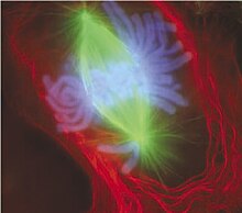
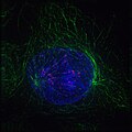
Real mitotic cells can be visualized through the microscope by staining them with fluorescent antibodies and dyes. These light micrographs are included below.
See also
References
- ^ De Souza CP, Osmani SA. (2007). "Mitosis, not just open or closed". Eukaryotic Cell 6 (9): 1521–7. doi:10.1128/EC.00178-07. PMID 17660363.
- ^ Maton A, Hopkins JJ, LaHart S, Quon Warner D, Wright M, Jill D. (1997). Cells: Building Blocks of Life. New Jersey: Prentice Hall. pp. 70–4. ISBN 0-13423476-6.
- ^ a b Lilly M, Duronio R. (2005). "New insights into cell cycle control from the Drosophila endocycle". Oncogene 24 (17): 2765–75. doi:10.1038/sj.onc.1208610. PMID 15838513.
- ^ a b Blow J, Tanaka T. (2005). "The chromosome cycle: coordinating replication and segregation. Second in the cycles review series". EMBO Rep 6 (11): 1028–34. doi:10.1038/sj.embor.7400557. PMID 16264427.
- ^ Zhou J, Yao J, Joshi H. (2002). "Attachment and tension in the spindle assembly checkpoint". Journal of Cell Science 115 (Pt 18): 3547–55. doi:10.1242/jcs.00029. PMID 12186941.
- ^ Nanninga N. (2001). "Cytokinesis in prokaryotes and eukaryotes: common principles and different solutions". Microbiology and Molecular Biology Reviews 65 (2): 319–33. doi:10.1128/MMBR.65.2.319-333.2001. PMID 11381104.
- ^ a b Lloyd C, Chan J. (2006). "Not so divided: the common basis of plant and animal cell division". Nature reviews. Molecular cell biology 7 (2): 147–52. doi:10.1038/nrm1831. PMID 16493420.
- ^ Raven et al., 2005, pp. 58–67.
- ^ Varmark H (2004). "Functional role of centrosomes in spindle assembly and organization". Journal of Cellular Biochemistry 91 (5): 904–14. doi:10.1002/jcb.20013. PMID 15034926.
- ^ Heywood P. (1978). "Ultrastructure of mitosis in the chloromonadophycean alga Vacuolaria virescens". Journal of Cell Science 31: 37–51. PMID 670329.
- ^ Ribeiro K, Pereira-Neves A, Benchimol M. (2002). "The mitotic spindle and associated membranes in the closed mitosis of trichomonads". Biology of the Cell 94 (3): 157–72. doi:10.1016/S0248-4900(02)01191-7. PMID 12206655.
- ^ Chan G, Liu S, Yen T. (2005). "Kinetochore structure and function". Trends in Cell Biology 15 (11): 589–98. doi:10.1016/j.tcb.2005.09.010. PMID 16214339.
- ^ a b Maiato H, DeLuca J, Salmon E, Earnshaw W. (2004). "The dynamic kinetochore-microtubule interface". Journal of Cell Science 117 (Pt 23): 5461–77. doi:10.1242/jcs.01536. PMID 15509863.
- ^ a b Winey M, Mamay C, O'Toole E, Mastronarde D, Giddings T, McDonald K, McIntosh J. (1995). "Three-dimensional ultrastructural analysis of the Saccharomyces cerevisiae mitotic spindle". Journal of Cell Biology 129 (6): 1601–15. doi:10.1083/jcb.129.6.1601. PMID 7790357.
- ^ Chan G, Yen T. (2003). "The mitotic checkpoint: a signaling pathway that allows a single unattached kinetochore to inhibit mitotic exit". Progress in Cell Cycle Research 5: 431–9. PMID 14593737.
- ^ Miller KR. (2000). "Anaphase". Biology (5 ed.). Pearson Prentice Hall. pp. 169–70. ISBN 978-0134362656.
- ^ Glotzer M. (2005). "The molecular requirements for cytokinesis". Science 307 (5716): 1735–9. doi:10.1126/science.1096896. PMID 15774750.
- ^ Albertson R, Riggs B, Sullivan W. (2005). "Membrane traffic: a driving force in cytokinesis". Trends in Cell Biology 15 (2): 92–101. doi:10.1016/j.tcb.2004.12.008. PMID 15695096.
- ^ Raven et al., 2005, pp. 64–7, 328–9.
- ^ Zhou G, Liu D, Liang C. (2005). "Memory mechanisms of active transcription during cell division". Bioessays 27 (12): 1239–45. doi:10.1002/bies.20327. PMID 16299763.
- ^ Draviam V, Xie S, Sorger P. (2004). "Chromosome segregation and genomic stability". Current Opinion in Genetics & Development 14 (2): 120–5. doi:10.1016/j.gde.2004.02.007. PMID 15196457.
- ^ Italiano JE, Shivdasani RA. (2003). "Megakaryocytes and beyond: the birth of platelets". Journal of Thrombosis and Haemostasis 1 (6): 1174–82. doi:10.1046/j.1538-7836.2003.00290.x. PMID 12871316.
Cited texts
- Raven PH, Evert RF, Eichhorn SE. (2005). Biology of Plants (7th ed.). New York: W.H. Freeman and Company Publishers. ISBN 0-7167-1007-2.
Further reading
- Morgan, David L. (2007). The cell cycle: principles of control. London: Published by New Science Press in association with Oxford University Press. ISBN 0-9539181-2-2.
- Alberts B, Johnson A, Lewis J, Raff M, Roberts K, and Walter P (2002). "Mitosis". Molecular Biology of the Cell. Garland Science. http://www.ncbi.nlm.nih.gov/books/bv.fcgi?highlight=mitosis&rid=mboc4.section.3349. Retrieved 2006-01-22.
- Campbell, N. and Reece, J. (December 2001). "The Cell Cycle". Biology (6th ed.). San Francisco: Benjamin Cummings/Addison-Wesley. pp. 217–224. ISBN 0-8053-6624-5.
- Cooper, G. (2000). "The Events of M Phase". The Cell: A Molecular Approach. Sinaeur Associates, Inc. http://www.ncbi.nlm.nih.gov/books/bv.fcgi?highlight=M%20Phase,Events&rid=cooper.section.2470. Retrieved 2006-01-22.
- Freeman, S (2002). "Cell Division". Biological Science. Upper Saddle River, NJ: Prentice Hall. pp. 155–174. ISBN 0-13-081923-9.
- Lodish H, Berk A, Zipursky L, Matsudaira P, Baltimore D, Darnell J (2000). "Overview of the Cell Cycle and Its Control". Molecular Cell Biology. W.H. Freeman. http://www.ncbi.nlm.nih.gov/books/bv.fcgi?highlight=Overview,Control,Cell+Cycle&rid=mcb.section.3463. Retrieved 2006-01-22.
External links
| Wikimedia Commons has media related to: Mitosis |
- Mitosis Animation.
- Mitosis (Flash Animation)
- Video of a live amphibian lung cell undergoing mitosis.
- A Flash animation comparing Mitosis and Meiosis
- Animation from the Life Sciences Dept of University of North Texas
- Studying Mitosis in Cultured Mammalian Cells
- General K-12 classroom resources for Mitosis
- CCO The Cell-Cycle Ontology
| |||||||||||||||||||||||||||||||
Personal tools
Namespaces
Variants
Print/export
Languages
- العربية
- বাংলা
- Bân-lâm-gú
- Bosanski
- Български
- Català
- Česky
- Dansk
- Deutsch
- Eesti
- Ελληνικά
- Español
- Esperanto
- Euskara
- فارسی
- Français
- Galego
- 한국어
- Հայերեն
- Hrvatski
- Ido
- Bahasa Indonesia
- Italiano
- עברית
- Kreyòl ayisyen
- Lietuvių
- Magyar
- Македонски
- Bahasa Melayu
- Nederlands
- 日本語
- Norsk (bokmål)
- Polski
- Português
- Русский
- Shqip
- Simple English
- Slovenčina
- Slovenščina
- Српски / Srpski
- Srpskohrvatski / Српскохрватски
- Suomi
- Svenska
- ไทย
- Türkçe
- Українська
- اردو
- Tiếng Việt
- 中文
- This page was last modified on 12 January 2011 at 07:57.
- Text is available under the Creative Commons Attribution-ShareAlike License; additional terms may apply. See Terms of Use for details. Wikipedia® is a registered trademark of the Wikimedia Foundation, Inc., a non-profit organization.
- Contact us
Mitosis
Mitosis: Labeled Diagram

Legend: Illustration of the process by which somatic cells multiply and divide.
Mitosis is a process of cell division which results in the production of two daughter cells from a single parent cell. The daughter cells are identical to one another and to the original parent cell.
In a typical animal cell, mitosis can be divided into four principals stages:
- Prophase: The chromatin, diffuse in interphase, condenses into chromosomes. Each chromosome has duplicated and now consists of two sister chromatids. At the end of prophase, the nuclear envelope breaks down into vesicles.
- Metaphase: The chromosomes align at the equitorial plate and are held in place by microtubules attached to the mitotic spindle and to part of the centromere.
- Anaphase: The centromeres divide. Sister chromatids separate and move toward the corresponding poles.
- Telophase: Daughter chromosomes arrive at the poles and the microtubules disappear. The condensed chromatin expands and the nuclear envelope reappears. The cytoplasm divides, the cell membrane pinches inward ultimately producing two daughter cells (phase: Cytokinesis).
Phases of Mitosis
5 Phases of Mitosis
Based on the changes occuring in the cell, five phases have been recognised inmitosis (1) Prophase (2) Metaphase (3) Anaphase (4) Telophase and (5) Cytokinesis
Prophase : This is the first stage of cell division. Before prophase starts, the chromosomes are in decondensed stage and appear as a diffuse network of filaments. During prophase, the chromosomes start condensing. As a result, the shape of the chromosomes becomes definite and distince. Each chromosome has two arms called chromatids. The two chromatids are joined together by centromere. While these changes are occurring, the nucleolus and rest of the structures in the nucleus (except chromosomes) disappear gradually. Meanwhile, the centrioles move to the opposite sides of nucleus. In plant cells, where centrioles are usually absent, they apear at this stage of cell division. From each centriole fine thread like fibres appear. These fibres are called spindle fibres. A centriole with spindle fibres is called an aster. The spindle fibres grow towards the centre of the cell.
Metaphase : Chromosomes now move to the centre of the cell and arrange themselves in a row. This look like a plate and is called equitorial plate. Some of the spindle fibres are attached to the centromere of each chromosome. Rest of the fibres grow towards opposite pole (or side) and join the centriole on the opposite side. Each chromosome is attached to fibres from the two opposite poles.
Anaphase: At this stage, spindle fibres pull the chromosomes to opposite ends. As a result, the centromere splits and are pulled apart along with chromatids. As a result, half the original number of chromosomes move to each of the two opposite poles.
Telophase : By the beginning of this phase, the chromosomes complete movement and will be at each end of the cell. Nuclear membrane, nucleolus and other nuclear structures reappear. Chromosomes begin decondensation process. By the end of Telophase, cell will be having two nuclei, one on each side. Spindle Fibres now disappear.
Cytokineses : A new membrane (and a cell wall in plant cells) is formed at the equatorial plate during this phase. This divides the cell into two daughter cells.

Conclusion to 5 Phases of Mitosis
Thus at the end of mitosis, two daughter cells which are exactly similar to the mother cell are formed. The first four stages of cell division involve changes in nucleus and division of nuclear components equally between daughter cells. Hence , these four phases (prophase, metaphase, anaphase and telophase ) of cell division are together called karyokinesis (division of nucleus). The last stage is called cytokinesis as the cytoplasm is the only component undergoing division in this stage of mitosis.
Explore Related Concepts
- Phases of mitosis
- Mitosis phase
- Mitosis phases
- Mitosis and meiosis phases
- 4 phases of mitosis
- Four phases of mitosis
- Mitosis diagram
- Mitosis images
- Anaphase of mitosis
- Mitosis anaphase
- Mitosis and Meiosis - A Comparison
- Four stages of mitosis
- Mitosis stages quiz
- Mitosis
- Differences between mitosis and meiosis
- Photochemical and Biosynthetic Phases
- Phase Constant in SHM
- Differences between Mitosis and Meiosis
- 5 stages of cell division
- Anaphase picture
- Binary fission animation
- Interphase
- Anaphase diagram
- Prophase
- Difference between Absorption and Adsorption
- Menstruation and Menstrual Cycle
- Population Growth
- Growth Curve
- Spermatogenesis
Cell Division
CELL DIVISION
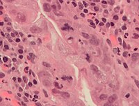
Cell division is the process by which new cells arise. A cell that is undergoing division is called a mitotic figure. The importance of learning to recognize mitotic figures should be emphasized. For example, the number of mitotic figures in a tissue provides an idea of the rate of turnover of cells in that tissue. Recognition of mitotic figures is useful in interpreting various kinds of pathology. For example, mitotic figures are usually absent in a benign tumor whereas they are usually numerous in a malignant tumor.
Types of cell division
1. Meiosis. In this type of cell division, the number of chromosomes is halved. The diploid number (46) is reduced to the haploid number (23) in the daughter cells. Meiosis is restricted to gametes (spermatocytes and oocytes)
2. Amitosis. Amitosis is often referred to as direct cell division. It is a type of cell division where the cell breaks into two parts by constriction of the nucleus and cytoplasm. There may be unequal distribution of the chromosomes. This type of cell division occurs in the placenta and in the surface cells of transitional epithelium.
3. Mitosis. This type of cell division is often called indirect cell division. It is characterized by an exact division and distribution of the chromosomal material between the two daughter cells. There is a division of the cytoplasm (cytokinesis) and a division of the nucleus (karyokinesis). It is the most common type of cell division.
Events prior to mitosis. These are events, which occur during interphase.
1. Replication of chromosomal DNA. Replication occurs at the molecular level and is not visible microscopically.
2. Growth of the cell.
Stages of mitosis. There are four stages: prophase, metaphase, anaphase, and telophase. Each stage merges imperceptibly into the next, making it impossible in some cases to recognize the end of one stage and the beginning of the next. It should be stressed that mitosis is a continuous process, which is arbitrarily divided into four stages. See Figure 9 below.
1. Prophase. Events include:
a. Chromosomes become visible. They are delicate coiled filaments at first, but later shorten and thicken.
b. Each chromosome divides longitudinally into two chromatids. The
chromatids, two of these constituting a chromosome, remain attached at some point along their length. This zone of attachment is called the centromere and may be located anywhere along the length of the two chromatids.
c. Centrioles migrate to opposite nuclear poles.
d. Nucleolus disappears. This event occurs in late prophase.
e. Nuclear membrane disappears. This event also occurs in late prophase.
f. Mitotic spindle appears. The mitotic spindle is a system of microtubules, which develop in the area between the two centrioles.
g. Kinetochore appears. The kinetochore is a small disc-like structure, which has been shown by electron microscopy to develop on each chromatid at the site of the centromere. Some of the microtubules of the mitotic spindle become attached to the kinetochore.

Figure 9: Phases of Mitosis. Taken from: Junqueira and Carneiro, Basic Histology, Text and Atlas, p. 60, Figure 3-15.
2. Metaphase. Events include:
a. Chromosomes become fully condensed and their centromeres aligned midway in the mitotic spindle. The centromeres occupy the area referred to as the equatorial or metaphase plate of the cell.
b. Mitotic spindle becomes fully developed. Two groups of microtubules form the spindle: chromosomal tubules and continuous tubules. The latter are not attached to the centriole at the other pole. Chromosomal tubules extend from the kinetochore of a chromatid to one of the centrioles. The kinetochore of the other chromatid of the chromosome is attached by chromosomal tubules to the opposite centriole. Thus, each chromosome has tubules from the other centriole attached to its other chromatid. Colchicine has been shown to arrest mitosis at the metaphase stage. It interferes with formation of the microtubules of the mitotic spindle, leaving the cell suspended in metaphase and unable to complete mitosis.
c. Disappearance of the nuclear envelope and nucleolus.
Colchicine is sometimes added to cultures of leukocytes whose mitotic activity has been stimulated with phytohemagglutinin, an extract from the red kidney bean. The cells are then spread on glass slides and stained. The chromosomes of these cells arrested in metaphase by colchicine can be studied and photographed. The photographs can be cut up, and the pairs of identical chromosomes can be matched to form a karyotype of the chromosomes of the individual from whom the cells were taken. The karyotype can be studied for abnormalities in number and form of the chromosomes.
3. Anaphase. Events include:
a. Centromeres split. The splitting may be due to the pull exerted on them by the microtubules as they move in opposite directions. Each chromatid is now called a chromosome.
b. Chromosomes move to opposite poles of the cell. In moving toward the cell pole, each chromosome travels with the kinetochore leading the way, the remainder of the chromosome appearing to be dragged behind. Anaphase is concluded when the two chromosomal masses have moved to opposite poles of the cell.
c. Centrioles divide.
4. Telophase. Events are reverse of prophase.
a. Chromosomes lose their identity. They straighten out into long thin strands, and they are so fine that they are beyond the resolution of the light microscope.
b. Nuclear membrane appears. It develops from cisternae of endoplasmic reticulum.
c. Nucleolus appears.
d. Organelles distributed between two daughter cells. They are distributed in approximately equal numbers.
Identification of mitotic figures in tissue sections
Although you should be familiar with the process of mitosis from previous courses, you should review the textbook material on mitosis before identifying mitotic figures in the class slides. Compare mitotic figures you see in the microscope with textbook illustrations. Frequently you will be asked to refer to your textbook as an aid to understanding the laboratory work and for purposes of orienting you in regard to unfamiliar tissues. You should therefore always bring your textbook to the laboratory.
You should learn to recognize cells in the various stages of cell division (mitosis) and develop an awareness of those region(s) of organs and tissues in which “mitotic figures" are most numerous. Study mitotic figures in the following slides.
Slide A1, Vicia faba, root tip, (Feulgen). This slide was prepared from a growing squash root, stained with the Feulgen technique, which is relatively specific for DNA, to show the various stages of mitosis. Individual chromosomes can be seen. Find prophase, metaphase, anaphase, and telophase stages in this slide. Try to recognize a mitotic figure with the low power objectives for careful study.
Slide A2, Ambystoma larva, epidermis, whole mount, (Fe-Hematoxylin). Skin from an
amphibian larva. You will have to focus up and down as the piece of tissue has numerous folds. You should find good examples of the stages of mitosis. The large, brown dendritic-appearing cells are subepithelial melanophores.
Slide B60, ileum, monkey, (H&E). “Mitotic figures” typical of tissue sections can be seen in the intestinal crypts (base of the finger-like projections or villi). Some, but not all, of the mitotic stages can be found.
Diagram of The Stage of Meiosis

| Diagram of the stages of meiosis: two stage division of a cell, producing gametes and halving the number of chromosomes in its nucleus. Prophase I: first phase of meiosis. Metaphase I: second phase of meiosis. Anaphase I: third phase of meiosis. Telophase I: fourth phase of meiosis. Prophase II: fifth phase of meiosis. Metaphase II: sixth phase of meiosis. Anaphase II: seventh phase of meiosis. Telophase II: eight phase of meiosis. Four cells: last phase of meiosis. |
Phases of Meiosis
The Phases of Meiosis
| |
| Meiosis begins with Interphase I. During this phase there is a duplication genetic material, DNA replication. Cells go from being 2N, 2C (N= chromosome content, C = DNA content) to 2N, 4C. Cells remain in this active phase 75% of the time. The chromatin remains in a nuclear envelope while a pair of centrioles lies inside a centrosome. During Prophase I, the chromatin condenses into chromosomes, the nuclear envelope disappears, and a spindle apparatus begins to form. Each chromosome consists of a pair of chromatids connected by a centromere. Cells are now 4N, 4C. The major occurrence in this phase is the coupling of these homologous chromosomes. Two double-stranded chromosomes form a four-stranded tetrad. In some cases, there is crossing-over of the two middle strands, at a site called the chiasma, such that there is genetic recombination. This process is extremely important for creating genetic diversity. In Metaphase I, the tetrads line up on the "equator" of the cell. The centrosome has replicated and one has moved to each pole. Microtubules that extend out of each centrosome attach to kinetochores in the center of each side of the tetrads that have lined up on the equator. | |
|
Anaphase I occurs as the microtubules pull the pairs of homologous chromatids toward each pole, as the tetrad is divided. The cell begins to lengthen. During Telophase I, the nuclear envelope begins to reform and nucleoli reappear. The cell begins to split, forming a cleavage furrow in the middle. In Cytokinesis I, the cells finally split, with one copy of each chromosome in each one. Each of the two resulting cells is now 2N, 2C.
| |
| Interkinesis has not replication, unlike the previous Interphase I and the interphase of mitosis. Prophase II, Metaphase II, Anaphase II, and Telophase II repeats the same steps as Prophase I-Telophase I, with half as much genetic material. Cytokinesis II is the final step of meiosis, where each cell splits into two daughter cells, for a total of four gametes, each with half the number of chromosomes. Each of the four resulting cells is 1N, 1C. (30) return to: The Process of Meiosis | |
Cell Division:Meiosis and sexual Reproduction
CELL DIVISION: MEIOSIS AND SEXUAL REPRODUCTION
Table of Contents
Meiosis | Ploidy | Life Cycles | Phases of Meiosis | Prophase I | Metaphase I
Anaphase I | Telophase I | Prophase II | Metaphase II | Anaphase II | Telophase II
Comparison of Mitosis and Meiosis | Gametogenesis | Links
Meiosis | Back to Top
Sexual reproduction occurs only in eukaryotes. During the formation of gametes, the number of chromosomes is reduced by half, and returned to the full amount when the two gametes fuse during fertilization.
Ploidy | Back to Top
Haploid and diploid are terms referring to the number of sets of chromosomes in a cell. Gregor Mendel determined his peas had two sets of alleles, one from each parent. Diploid organisms are those with two (di) sets. Human beings (except for their gametes), most animals and many plants are diploid. We abbreviate diploid as 2n. Ploidy is a term referring to the number of sets of chromosomes. Haploid organisms/cells have only one set of chromosomes, abbreviated as n. Organisms with more than two sets of chromosomes are termed polyploid. Chromosomes that carry the same genes are termed homologous chromosomes. The alleles on homologous chromosomes may differ, as in the case of heterozygous individuals. Organisms (normally) receive one set of homologous chromosomes from each parent.
Meiosis is a special type of nuclear division which segregates one copy of each homologous chromosome into each new "gamete". Mitosis maintains the cell's original ploidy level (for example, one diploid 2n cell producing two diploid 2n cells; one haploid n cell producing two haploid n cells; etc.). Meiosis, on the other hand, reduces the number of sets of chromosomes by half, so that when gametic recombination (fertilization) occurs the ploidy of the parents will be reestablished.
Most cells in the human body are produced by mitosis. These are the somatic (or vegetative) line cells. Cells that become gametes are referred to as germ line cells. The vast majority of cell divisions in the human body are mitotic, with meiosis being restricted to the gonads.
Life Cycles | Back to Top
Life cycles are a diagrammatic representation of the events in the organism's development and reproduction. When interpreting life cycles, pay close attention to the ploidy level of particular parts of the cycle and where in the life cycle meiosis occurs. For example, animal life cycles have a dominant diploid phase, with the gametic (haploid) phase being a relative few cells. Most of the cells in your body are diploid, germ line diploid cells will undergo meiosis to produce gametes, with fertilization closely following meiosis.
Plant life cycles have two sequential phases that are termed alternation of generations. The sporophyte phase is "diploid", and is that part of the life cycle in which meiosis occurs. However, many plant species are thought to arise by polyploidy, and the use of "diploid" in the last sentence was meant to indicate that the greater number of chromosome sets occur in this phase. The gametophyte phase is "haploid", and is the part of the life cycle in which gametes are produced (by mitosis of haploid cells). In flowering plants (angiosperms) the multicelled visible plant (leaf, stem, etc.) is sporophyte, while pollen and ovaries contain the male and female gametophytes, respectively. Plant life cycles differ from animal ones by adding a phase (the haploid gametophyte) after meiosis and before the production of gametes.
Many protists and fungi have a haploid dominated life cycle. The dominant phase is haploid, while the diploid phase is only a few cells (often only the single celled zygote, as in Chlamydomonas ). Many protists reproduce by mitosis until their environment deteriorates, then they undergo sexual reproduction to produce a resting zygotic cyst.
Phases of Meiosis | Back to Top
Two successive nuclear divisions occur, Meiosis I (Reduction) and Meiosis II (Division). Meiosis produces 4 haploid cells. Mitosis produces 2 diploid cells. The old name for meiosis was reduction/ division. Meiosis I reduces the ploidy level from 2n to n (reduction) while Meiosis II divides the remaining set of chromosomes in a mitosis-like process (division). Most of the differences between the processes occur during Meiosis I.

The above image is from http://www.biology.uc.edu/vgenetic/meiosis/
Prophase I | Back to Top
Prophase I has a unique event -- the pairing (by an as yet undiscovered mechanism) of homologous chromosomes. Synapsis is the process of linking of the replicated homologous chromosomes. The resulting chromosome is termed a tetrad, being composed of two chromatids from each chromosome, forming a thick (4-strand) structure. Crossing-over may occur at this point. During crossing-over chromatids break and may be reattached to a different homologous chromosome.
The alleles on this tetrad:
A B C D E F G
A B C D E F G
a b c d e f g
a b c d e f g
will produce the following chromosomes if there is a crossing-over event between the 2nd and 3rd chromosomes from the top:
A B C D E F G
A B c d e f g
a b C D E F G
a b c d e f g
Thus, instead of producing only two types of chromosome (all capital or all lower case), four different chromosomes are produced. This doubles the variability of gamete genotypes. The occurrence of a crossing-over is indicated by a special structure, a chiasma (plural chiasmata) since the recombined inner alleles will align more with others of the same type (e.g. a with a, B with B). Near the end of Prophase I, the homologous chromosomes begin to separate slightly, although they remain attached at chiasmata.

Crossing-over between homologous chromosomes produces chromosomes with new associations of genes and alleles. Image from Purves et al., Life: The Science of Biology, 4th Edition, by Sinauer Associates (www.sinauer.com) and WH Freeman (www.whfreeman.com), used with permission.
Events of Prophase I (save for synapsis and crossing over) are similar to those in Prophase of mitosis: chromatin condenses into chromosomes, the nucleolus dissolves, nuclear membrane is disassembled, and the spindle apparatus forms.


Major events in Prophase I. Image from Purves et al., Life: The Science of Biology, 4th Edition, by Sinauer Associates (www.sinauer.com) and WH Freeman (www.whfreeman.com), used with permission.
Metaphase I | Back to Top
Metaphase I is when tetrads line-up along the equator of the spindle. Spindle fibers attach to the centromere region of each homologous chromosome pair. Other metaphase events as in mitosis.
Anaphase I | Back to Top
Anaphase I is when the tetrads separate, and are drawn to opposite poles by the spindle fibers. The centromeres in Anaphase I remain intact.

Events in prophase and metaphse I. Image from Purves et al., Life: The Science of Biology, 4th Edition, by Sinauer Associates (www.sinauer.com) and WH Freeman (www.whfreeman.com), used with permission.
Telophase I | Back to Top
Telophase I is similar to Telophase of mitosis, except that only one set of (replicated) chromosomes is in each "cell". Depending on species, new nuclear envelopes may or may not form. Some animal cells may have division of the centrioles during this phase.

The events of Telophase I. Image from Purves et al., Life: The Science of Biology, 4th Edition, by Sinauer Associates (www.sinauer.com) and WH Freeman (www.whfreeman.com), used with permission.
Prophase II | Back to Top
During Prophase II, nuclear envelopes (if they formed during Telophase I) dissolve, and spindle fibers reform. All else is as in Prophase of mitosis. Indeed Meiosis II is very similar to mitosis.

The events of Prophase II. Image from Purves et al., Life: The Science of Biology, 4th Edition, by Sinauer Associates (www.sinauer.com) and WH Freeman (www.whfreeman.com), used with permission.
Metaphase II | Back to Top
Metaphase II is similar to mitosis, with spindles moving chromosomes into equatorial area and attaching to the opposite sides of the centromeres in the kinetochore region.
Anaphase II | Back to Top
During Anaphase II, the centromeres split and the former chromatids (now chromosomes) are segregated into opposite sides of the cell.

The events of Metaphase II and Anaphase II. Image from Purves et al., Life: The Science of Biology, 4th Edition, by Sinauer Associates (www.sinauer.com) and WH Freeman (www.whfreeman.com), used with permission.
Telophase II | Back to Top
Telophase II is identical to Telophase of mitosis. Cytokinesis separates the cells.

The events of Telophase II. Image from Purves et al., Life: The Science of Biology, 4th Edition, by Sinauer Associates (www.sinauer.com) and WH Freeman (www.whfreeman.com), used with permission.
Comparison of Mitosis and Meiosis | Back to Top
Mitosis maintains ploidy level, while meiosis reduces it. Meiosis may be considered a reduction phase followed by a slightly altered mitosis. Meiosis occurs in a relative few cells of a multicellular organism, while mitosis is more common.


Comparison of the events in Mitosis and Meiosis. Images from Purves et al., Life: The Science of Biology, 4th Edition, by Sinauer Associates (www.sinauer.com) and WH Freeman (www.whfreeman.com), used with permission.
Gametogenesis | Back to Top
Gametogenesis is the process of forming gametes (by definition haploid, n) from diploid cells of the germ line. Spermatogenesis is the process of forming sperm cells by meiosis (in animals, by mitosis in plants) in specialized organs known as gonads (in males these are termed testes). After division the cells undergo differentiation to become sperm cells. Oogenesis is the process of forming an ovum (egg) by meiosis (in animals, by mitosis in the gametophyte in plants) in specialized gonads known as ovaries. Whereas in spermatogenesis all 4 meiotic products develop into gametes, oogenesis places most of the cytoplasm into the large egg. The other cells, the polar bodies, do not develop. This all the cytoplasm and organelles go into the egg. Human males produce 200,000,000 sperm per day, while the female produces one egg (usually) each menstrual cycle.


Gametogenesis. Images from Purves et al., Life: The Science of Biology, 4th Edition, by Sinauer Associates (www.sinauer.com) and WH Freeman (www.whfreeman.com), used with permission.
Spermatogenesis
Sperm production begins at puberty at continues throughout life, with several hundred million sperm being produced each day. Once sperm form they move into the epididymis, where they mature and are stored.

Human Sperm (SEM x5,785). This image is copyright Dennis Kunkel at www.DennisKunkel.com, used with permission.
Oogenesis
The ovary contains many follicles composed of a developing egg surrounded by an outer layer of follicle cells. Each egg begins oogenesis as a primary oocyte. At birth each female carries a lifetime supply of developing oocytes, each of which is in Prophase I. A developing egg (secondary oocyte) is released each month from puberty until menopause, a total of 400-500 eggs.

Oogenesis. The above image is from http://www.grad.ttuhsc.edu/courses/histo/notes/female.html.
Links | Back to Top
- Access Excellence page on Mitosis
- Cell Division and the Cell Cycle (University of Alberta): Similar to this page, but with its own glossary and questions.
- Amoeba Proteus Mitosis Small photomicrographs of protistan mitosis.
- Animated Meiosis Yale University, a simplified series of cartoons about meiosis.
- Meiosis Tutorial North Carolina State University, animations and 3-D graphics.
- McGill University Mitosis Page Quality site, with photos and downloadable animation and video.
- Virtual Meiosis University of Cincinnati, Animated GIF and text/images to explain meiosis.
Text ©1992, 1994, 1997, 1998, 2000, 2001, 2007, by M.J. Farabee, all rights reserved. Use for educational purposes is encouraged.
Back to Table of Contents | Mitosis Page
Email: mj.farabee@emcmail.maricopa.edu![]()
Last modified: Tuesday May 18 2010
The URL of this page is: www.emc.maricopa.edu/faculty/farabee/BIOBK/BioBookmeiosis.html
Difference Between Mitosis and Meiosis
Mitosis vs Meiosis
Meiosis and Mitosis describe cell division in eukaryotic cells when the chromosome separates.
In mitosis chromosomes separates and form into two identical sets of daughter nuclei, and it is followed by cytokinesis (division of cytoplasm). Basically, in mitosis the mother cell divides into two daughter cells which are genetically identical to each other and to the parent cell.
Phases of mitosis include:

1. Interface -where cell prepares for cell division and it also includes three other phases such as G1 (growth), S (synthesis), and G2 (second gap) 2. Prophase – formation of centrosomes, condensation of chromatin 3. Prometaphase- degradation of the nuclear membrane, attachment of microtubules to kinetochores 4. Metaphase- alignment of chromosomes at the metaphase plate 5. Early anaphase- shortening of kinetochore microtubules 6. Telophase- de-condensation of chromosomes and surrounded by nuclear membranes, formation of cleavage furrow. 7. Cytokinesis- division of cytoplasm
Meiosis is a reductional cell division where the number of chromosomes is divided into half. Gametes formations occur in animal cell and meiosis is necessary for sexual reproduction which occurs in eukaryotes. Meiosis influence stable sexual reproduction by halving of ploidy or chromosome count. Without meiosis the fertilization would result in zygote with twice the number of the parent.
Phases of meiosis include:
 1. Meiosis I – separation of homologous chromosomes and production of two haploid cells (23 chromosomes, N in humans)
2. Prophase I – pairing of homologous chromosome pair and recombination (crossing over) occurs
3. Metaphase I – Homologous pairs move along the metaphase plate, kinetochore microtubules from both centrioles attach to the homologous chromosomes align along an equatorial plane.
4. Anaphase I – shortening of microtubules, pulling of chromosomes toward opposing poles, forming two haploid sets
5. Telophase I – arrival of chromosomes to the poles with each daughter cell containing half the number of chromosomes
6. Meiosis II – second part of the meiotic process with the production of four haploid cells from the two haploid cells
Read more: Difference Between Mitosis and Meiosis | Difference Between | Mitosis vs Meiosis http://www.differencebetween.net/science/difference-between-mitosis-and-meiosis/#ixzz19NhuSiT6


